

Ask someone to picture brain surgery, and they’ll usually describe something dramatic: a shaved head, a long operation, a very uncertain recovery. Some of that picture is outdated.
Operating rooms today look different. Doctors practice with the help of 3D models, monitor the tools on a monitor, and at times operate on a tumor without making a single incision on the skull using the nasal route. If you want to find a neurosurgeon in Ghaziabad or a neurosurgeon doctor near Ghaziabad, it is important to know what the technology can do and cannot do.
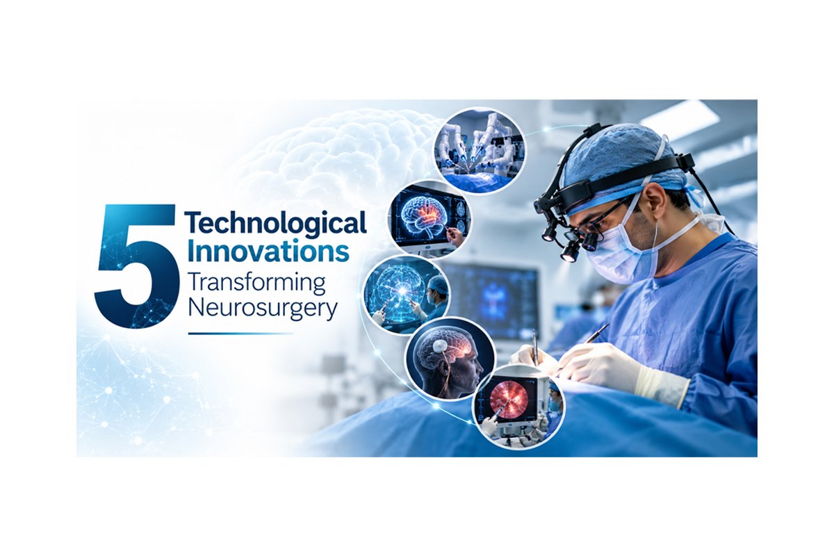
The following are five significant changes. This is only for general information, not personal health advice. Your scans and symptoms decide what’s right for you.
1. Neuronavigation: The GPS of Surgery
Prior to surgery, the patient is scanned using either a CT scan or an MRI scan. While the operation is in progress, the navigation system uses this scan to pinpoint the location on the body part of the patient being operated on. If the surgeon places the tip of an instrument at a certain spot, the computer screen will show the location on the scan, usually with an accuracy of one or two millimeters.
Why is that important? Because the brain is very congested and a tumor may be situated only a few millimeters away from the site responsible for controlling speech or movement, and nothing much resembles a map in terms of appearance.
Scanning in the middle of surgery
Others have gone a step further by performing intra-operative MRI. The MRI machine is inside the operating theatre, allowing the medical team to determine if there is any leftover tumor during the operation itself. This is particularly important as the brain will move slightly once the skull is opened, making the pre-operative MRI less accurate. Research involving glioma operations has shown that this technique leads to greater resection in selected cases. But the equipment is costly, and plenty of good hospitals don’t have it.
2. Robotic Assistance (The Surgeon Still Drives)
Let’s clear up one thing. Neurosurgical robots don’t operate by themselves. The surgeon plans the case and controls every step. What the robot adds is a very steady arm that holds a guide at the planned angle, something human hands can’t do for long without tiring.
You’ll see this most in three areas:
- Spine surgery, to place screws in the vertebrae along a pre-planned path
- Epilepsy surgery, to position thin electrodes that map where seizures start
- Deep brain stimulation, to reach tiny targets for conditions like Parkinson’s disease
Reported accuracy is good, but outcomes still depend on the team’s skill. A robot makes a trained surgeon more precise. It doesn’t turn an untrained one into a trained one.
3. Endoscopic and Minimally Invasive Techniques
Picture a camera as thin as a pencil, with tiny tools alongside it. That’s the basic idea of neuroendoscopy.
Pituitary tumours are the classic example. Most of them can be extracted via the nostril and sinus cavity behind the nostril, which makes no need for any incision to be made in the head. Some cases of hydrocephalus, a condition in which there is an accumulation of fluid within the brain, may be dealt with using a process known as endoscopic third ventriculostomy.
The spine has its own version. Endoscopic and tubular methods can treat some herniated discs through a cut of a centimetre or so, which usually means less muscle damage and a quicker return home.
One caution, though. Being “minimally invasive” may seem better, but it is not necessarily the case.In some cases, there are instances wherein surgery is necessary for your own safety. Your doctor can tell you which method of surgery suits you better.
4. Neuromonitoring and Glowing Tumours
Two tools work quietly in the background of difficult operations.
This monitoring system monitors the activity of the nerves and brain by detecting their electrical activity during the patient’s general anesthesia. If the activity drops, the monitoring will alert the surgical team immediately so that they can react. It’s widely used in spine deformity surgery and in operations close to the facial nerve or to motor areas of the brain.
Fluorescence-guided surgery is the one people find strangest. In the case of high-grade gliomas, 5-ALA is consumed by the patient before surgery. The tumour becomes fluorescent pink under specific light, whereas the normal brain does not fluoresce, making the demarcation line clear. Clinical trials showed that this helped surgeons remove more of the tumour in suitable cases.
And then there is awake surgery, where the patient is intentionally left awake during the procedure in order to be able to conduct certain tests on their speech or movement capabilities during the surgery process. It sounds terrifying, but it is thoroughly thought out and performed in specific cases only.
5. 3D Planning, Virtual Reality and Artificial Intelligence Imaging
In the past, surgeons had to make do with plain films and their imagination. They can now convert the scan into 3D format, visualize the tumor and its blood vessels at once, and practice their technique in virtual reality. Virtual and augmented reality are also changing training, because young surgeons can practise without any risk to patients. High-definition 3D microscopes and exoscopes give the whole team the same clear view of delicate work.
Artificial intelligence is arriving too, mostly in imaging. The software can assist in detecting any bleeding that may be present in the brain CT scan or even highlighting a tumor to aid with treatment planning. This is like another set of eyes. However, the interpretation of the scan and the decision are left to the physician, and this area advances rapidly enough to require some skepticism.
The Hospital Systems Nobody Talks About
None of this works if the right implant isn’t in the building. On the day of surgery, screws, shunts, plates, and disposable equipment should be readily available and traceable after the procedure is over. It is possible for hospitals running a good ordering process to correlate stocks with operating schedules. And reliable logistics tracking means any implant can be traced to its batch if a safety notice ever comes out. It’s not exciting, but it’s part of safe care.
Signs You Shouldn’t Wait
Some symptoms need urgent care at the nearest hospital, not a scheduled appointment:
- A sudden, severe headache unlike any before
- Weakness, numbness, or a drooping face on one side
- Trouble speaking or understanding speech
- A first-time seizure
- A head injury followed by repeated vomiting, drowsiness, or confusion
- Back pain with leg weakness, or loss of bladder or bowel control
The Final Word
Precision and gentle procedures have become a part of neurosurgery due to navigation, robotics, endoscopy, neuromonitoring, and 3D planning. Nevertheless, technology alone is not enough; the result depends on the correct diagnosis, counseling, teamwork, and proper monitoring.
When choosing a neurosurgeon in Ghaziabad, pay attention to such aspects as registration, communication, and a treatment plan that is suitable for your case. Don’t forget about your medical reports, and write down all your questions.
Disclaimer: This article is for general information only and doesn’t replace consultation with a registered medical practitioner. Availability of any technology varies by hospital.
- Tags:
- Healthcare